3.6.2. Saliva and food ingestion
Salivary secretions dilute the ingested food and adjust its pH and ionic content. The saliva often contains digestive enzymes and, in blood-feeding insects, anticoagulants and thinning agents are present. In insects with extra-intestinal digestion, such as predatory Hemiptera, digestive enzymes are exported into the food and the resulting liquid is ingested. Most Hemiptera produce an alkaline watery saliva that is a vehicle for enzymes (either digestive or lytic), and a proteinaceous solidifying saliva that either forms a complete sheath around the mouthparts (stylets) as they pierce and penetrate the food or just a securing flange at the point of entry (section 11.2.3, Fig. 11.4c). Stylet-sheath feeding is characteristic of phloem- and xylem-feeding Hemiptera, such as aphids, scale insects (coccoids), and spittle bugs, which leave visible tracks formed of exuded solidifying saliva in the plant tissue on which they have fed. The sheath may function to guide the stylets, prevent loss of fluid from damaged cells, and/or absorb necrosis-inducing compounds to reduce defensive reaction by the plant. By comparison, a macerate-and-flush strategy is typical of Heteroptera, such as mirids and coreids. These insects disrupt the tissues of plants or other insects by thrusting of the stylets and/or by addition of salivary enzymes. The macerated and/or partly digested food is “flushed out” with saliva and ingested by sucking.
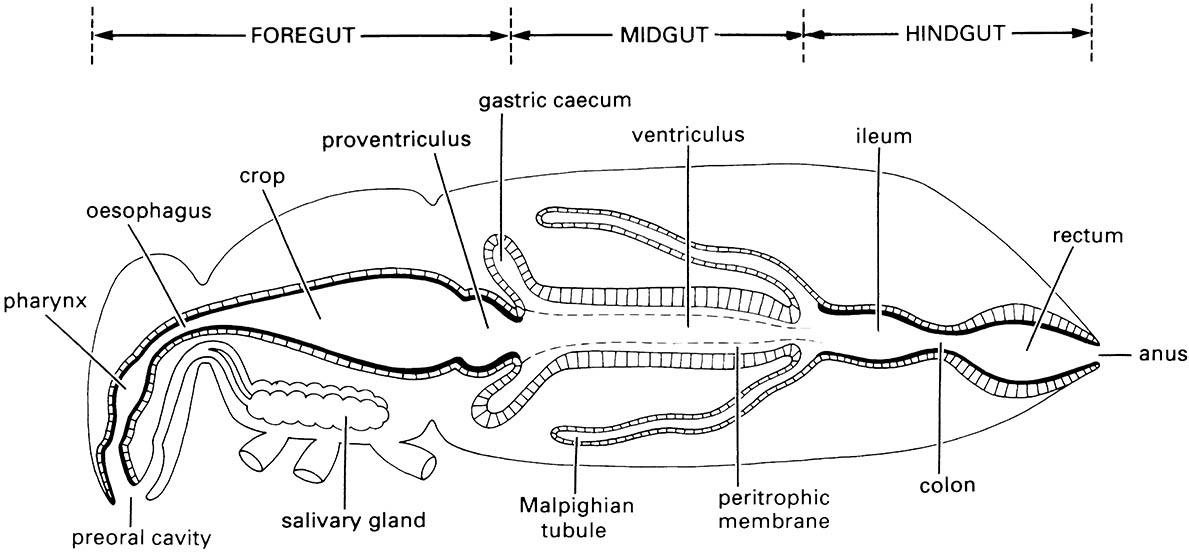
In fluid-feeding insects, prominent dilator muscles attach to the walls of the pharynx and/or the preoral cavity (cibarium) to form a pump (Fig. 3.14b), although most other insects have some sort of pharyngeal pump (Fig. 3.14a) for drinking and air intake to facilitate cuticle expansion during a molt.
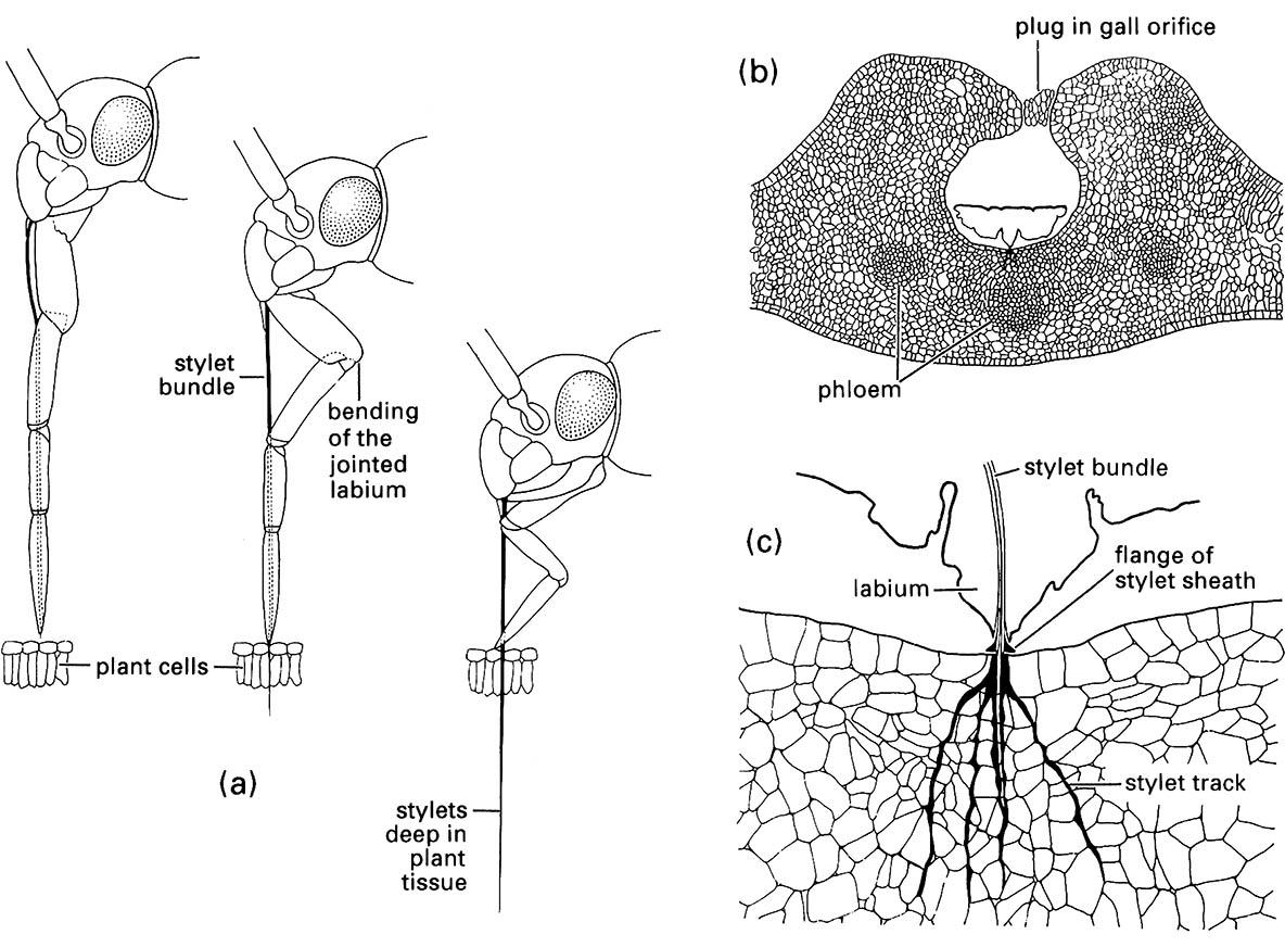
(a) penetration of plant tissue by a mirid bug showing bending of the labium as the stylets enter the plant; (b) transverse section through a eucalypt leaf gall containing a feeding nymph of a scale insect, Apiomorpha (Eriococcidae); (c) enlargement of the feeding site of (b) showing multiple stylet tracks (formed of solidifying saliva) resulting from probing of the parenchyma. ((a) After Poisson 1951)
Musculature of the mouthparts and the (a) pharyngeal or (b) cibarial pump are indicated but not fully labeled. Contraction of the respective dilator muscles causes dilation of the pharynx or cibarium and fluid is drawn into the pump chamber. Relaxation of these muscles results in elastic return of the pharynx or cibarial walls and expels food upwards into the oesophagus. (After Snodgrass 1935)

