3.4.2. Circulation
Circulation in insects is maintained mostly by a system of muscular pumps moving hemolymph through compartments separated by fibromuscular septa or membranes. The main pump is the pulsatile dorsal vessel. The anterior part may be called the aorta and the posterior part may be called the heart, but the two terms are inconsistently applied. The dorsal vessel is a simple tube, generally composed of one layer of myocardial cells and with segmentally arranged openings, or ostia. The lateral ostia typically permit the one-way flow of hemolymph into the dorsal vessel as a result of valves that prevent backflow. In many insects there also are more ventral ostia that permit hemolymph to flow out of the dorsal vessel, probably to supply adjacent active muscles. There may be up to three pairs of thoracic ostia and nine pairs of abdominal ostia, although there is an evolutionary tendency towards reduction in number of ostia.
The dorsal vessel lies in a compartment, the pericardial sinus, above a dorsal diaphragm (a fibromuscular septum — a separating membrane) formed of connective tissue and segmental pairs of alary muscles. The alary muscles support the dorsal vessel but their contractions do not affect heartbeat. Hemolymph enters the pericardial sinus via segmental openings in the diaphragm and/or at the posterior border and then moves into the dorsal vessel via the ostia during a muscular relaxation phase. Waves of contraction, which normally start at the posterior end of the body, pump the hemolymph forwards in the dorsal vessel and out via the aorta into the head. Next, the appendages of the head and thorax are supplied with hemolymph as it circulates posteroventrally and eventually returns to the pericardial sinus and the dorsal vessel. A generalized pattern of hemolymph circulation in the body is shown in Fig. 3.9a; however, in adult insects there also may be a periodic reversal of hemolymph flow in the dorsal vessel (from thorax posteriorly) as part of normal circulatory regulation.
Another important component of the circulation of many insects is the ventral diaphragm (Fig. 3.9b) — a fibromuscular septum that lies in the floor of the body cavity and is associated with the ventral nerve cord. Circulation of the hemolymph is aided by active peristaltic contractions of the ventral diaphragm, which direct the hemolymph backwards and laterally in the perineural sinus below the diaphragm. Hemolymph flow from the thorax to the abdomen also may be dependent, at least partially, on expansion of the abdomen, thus “sucking” hemolymph posteriorly. Hemolymph movements are especially important in insects that use the circulation in thermoregulation (some Odonata, Diptera, Lepidoptera, and Hymenoptera). Another function of the diaphragm may be to facilitate rapid exchange of chemicals between the ventral nerve cord and the hemolymph by either actively moving the hemolymph and/or moving the cord itself.
Hemolymph generally is circulated to appendages unidirectionally by various tubes, septa, valves, and pumps (Fig. 3.9c). The muscular pumps are termed accessory pulsatile organs and occur at the base of the antennae, at the base of the wings, and sometimes in the legs. Furthermore, the antennal pulsatile organs may release neurohormones that are carried to the antennal lumen to influence the sensory neurons. Wings have a definite but variable circulation, although it may be apparent only in the young adult. At least in some Lepidoptera, circulation in the wing occurs by the reciprocal movement of hemolymph (in the wing vein sinuses) and air (within the elastic wing tracheae) into and from the wing, brought about by pulsatile organ activity, reversals of heartbeat, and tracheal volume changes.
The insect circulatory system displays an impressive degree of synchronization between the activities of the dorsal vessel, fibromuscular diaphragms, and accessory pumps, mediated by both nervous and neurohormonal regulation. The physiological regulation of many body functions by the neurosecretory system occurs via neurohormones transported in the hemolymph.
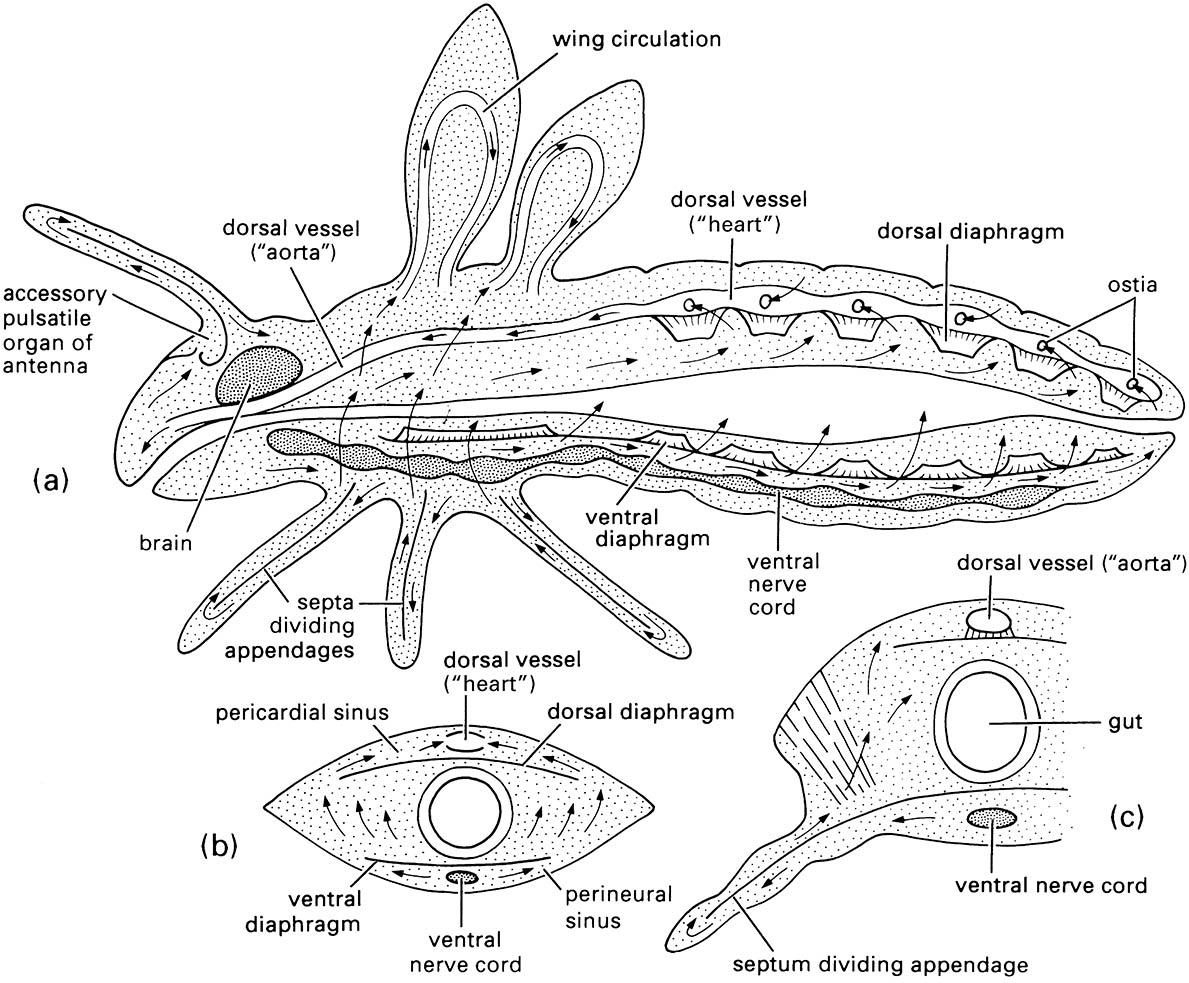
(a) longitudinal section through body; (b) transverse section of the abdomen; (c) transverse section of the thorax. Arrows indicate directions of hemolymph flow. (After Wigglesworth 1972)

