3.6.4. The fat body
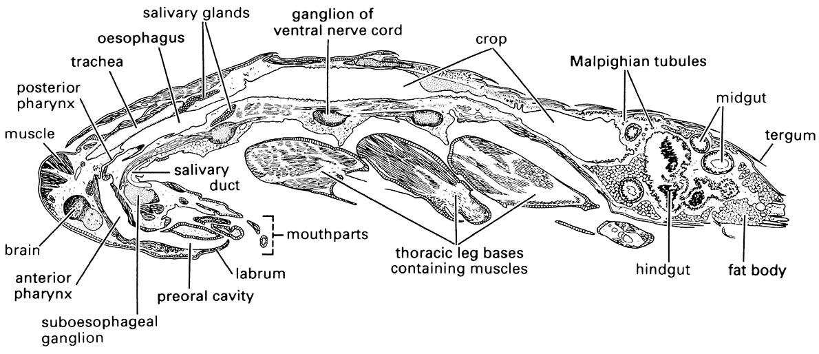
In many insects, especially the larvae of holometabol- ous groups, fat body tissue is a conspicuous component of the internal anatomy (Figs. 3.7 & 3.15). Typically, it forms a white or yellow tissue formed of loose sheets, ribbons, or lobes of cells lying in the hemocoel. The structure of this organ is ill-defined and taxonomically variable, but often caterpillars and other larvae have a peripheral layer of fat body beneath the cuticle and a central layer around the gut. The fat body is an organ of multiple metabolic functions, including: the metabolism of carbohydrates, lipids, and nitrogenous com- pounds; the storage of glycogen, fat, and protein; the synthesis and regulation of blood sugar; and the synthesis of major hemolymph proteins (such as hemoglobins, vitellogenins for yolk formation, and storage proteins). Fat body cells can switch their activities in response to nutritional and hormonal signals to supply the requirements of insect growth, metamorphosis, and reproduction. For example, specific storage proteins are synthesized by the fat body during the final larval instar of holometabolous insects and accumulate in the hemolymph to be used during metamorphosis as a source of amino acids for the synthesis of proteins during pupation. Calliphorin, a hemolymph storage protein synthesized in the fat body of larval blow flies (Diptera: Calliphoridae: Calliphora), may form about 75% of the hemolymph protein of a late-instar maggot, or about 7 mg; the amount of calliphorin falls to around 3 mg at the time of pupariation and to 0.03 mg after emergence of the adult fly. The production and deposition of proteins specifically for amino acid storage is a feature that insects share with seed plants but not with vertebrates. Humans, for example, excrete any dietary amino acids that are in excess of immediate needs.
The principal cell type found in the fat body is the trophocyte (or adipocyte), which is responsible for most of the above metabolic and storage functions. Visible differences in the extent of the fat body in different individuals of the same insect species reflect the amount of material stored in the trophocytes; little body fat indicates either active tissue construction or starvation. Two other cell types — urocytes and mycetocytes (also called bacteriocytes) — may occur in the fat body of some insect groups. Urocytes temporarily store spherules of urates, including uric acid, one of the nitrogenous wastes of insects. Amongst studied cockroaches, rather than being permanent stores of excreted waste uric acid (storage excretion), urocytes recycle urate nitrogen, perhaps with assistance of mycetocyte bacteria. Mycetocytes (bacteriocytes) contain symbiotic microorganisms and are scattered through the fat body of cockroaches or contained within special organs, sometimes surrounded by fat body. These bacteria-like symbionts appear important in insect nutrition.
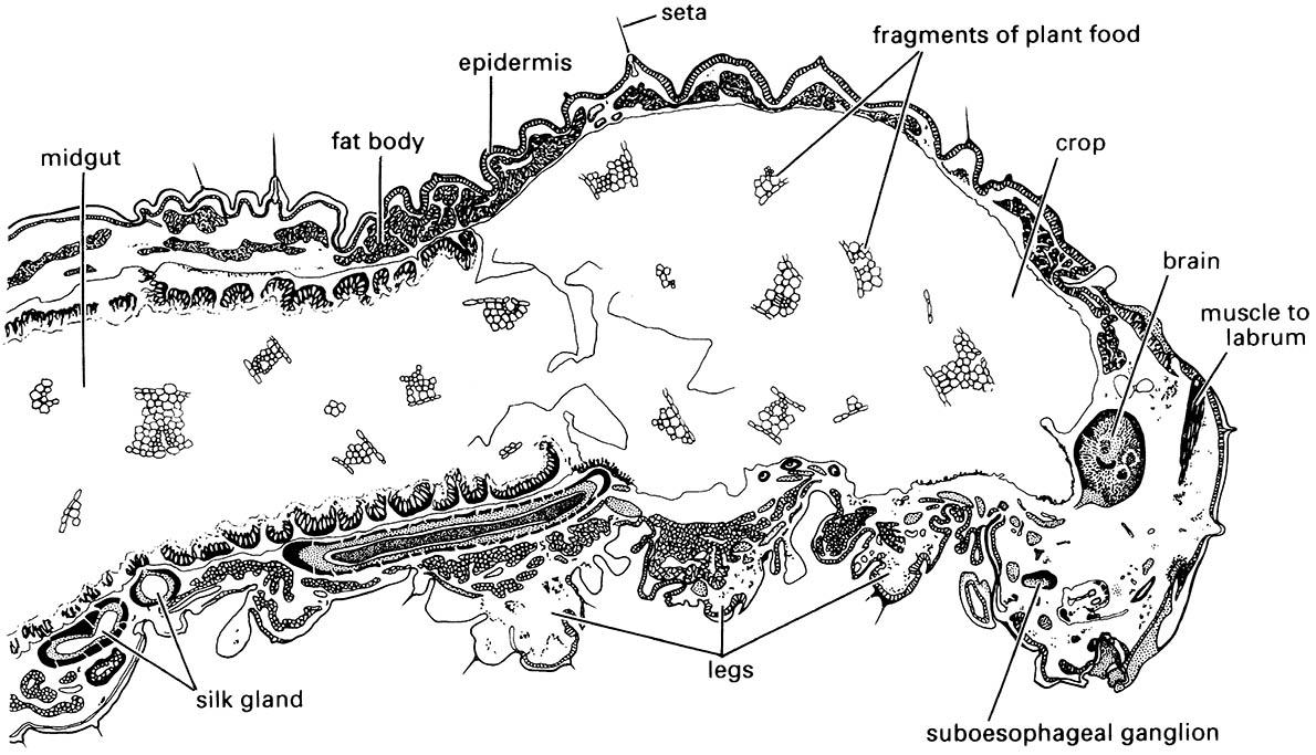
Note the thickened epidermal layer lining the midgut.

