3.7.1. The Malpighian tubules and rectum
The main organs of excretion and osmoregulation in insects are the Malpighian tubules acting in concert with the rectum and/or ileum (Fig. 3.17). Malpighian tubules are outgrowths of the alimentary canal and consist of long thin tubules (Fig. 3.1) formed of a single layer of cells surrounding a blind-ending lumen. They range in number from as few as two in most scale insects (coccoids) to over 200 in large locusts. Generally they are free, waving around in the hemolymph, where they filter out solutes. Only aphids lack Malpighian tubules. The vignette for this chapter shows the gut of Locusta, but with only a few of the many Malpighian tubules depicted. Similar structures are believed to have arisen convergently in different arthropod groups, such as myriapods and arachnids, in response to the physiological stresses of life on dry land. Traditionally, insect Malpighian tubules are considered to belong to the hindgut and be ectodermal in origin. Their position marks the junction of the midgut and the cuticle-lined hindgut.
The anterior hindgut is called the ileum, the generally narrower middle portion is the colon, and the expanded posterior section is the rectum (Fig. 3.13). In many terrestrial insects the rectum is the only site of water and solute resorption from the excreta, but in other insects, for example the desert locust Schistocerca gregaria (Orthoptera: Acrididae), the ileum makes some contribution to osmoregulation. In a few insects, such as the cockroach Periplaneta americana (Blattodea: Blattidae), even the colon may be a potential site of some fluid absorption. The resorptive role of the rectum (and sometimes the anterior hindgut) is indicated by its anatomy. In most insects, specific parts of the rectal epithelium are thickened to form rectal pads or papillae composed of aggregations of columnar cells; typically there are six pads arranged longitudinally, but there may be fewer pads or many papillate ones.
The general picture of insect excretory processes outlined here is applicable to most freshwater species and to the adults of many terrestrial species. The Malpighian tubules produce a filtrate (the primary urine) which is isosmotic but ionically dissimilar to the hemolymph, and then the hindgut, especially the rectum, selectively reabsorbs water and certain solutes but eliminates others (Fig. 3.17). Details of Malpighian tubule and rectal structure and of filtration and absorption mechanisms differ between taxa, in relation to both taxonomic position and dietary composition (Box 3.4 gives an example of one type of specialization — cryptonephric systems), but the excretory system of the desert locust S. gregaria (Fig. 3.18) exemplifies the general structure and principles of insect excretion. The Malpighian tubules of the locust produce an isosmotic filtrate of the hemolymph, which is high in K+, low in Na+, and has Cl- as the major anion. The active transport of ions, especially K+, into the tubule lumen generates an osmotic pressure gradient so that water passively follows (Fig. 3.18a).
Sugars and most amino acids also are filtered passively from the hemolymph (probably via junctions between the tubule cells), whereas the amino acid proline (later used as an energy source by the rectal cells) and non-metabolizable and toxic organic compounds are transported actively into the tubule lumen. Sugars, such as sucrose and trehalose, are resorbed from the lumen and returned to the hemolymph. The continuous secretory activity of each Malpighian tubule leads to a flow of primary urine from its lumen towards and into the gut. In the rectum, the urine is modified by removal of solutes and water to maintain fluid and ionic homeostasis of the locust’s body (Fig. 3.18b). Specialized cells in the rectal pads carry out active recovery of Cl- under hormonal stimulation. This pumping of Cl- generates electrical and osmotic gradients that lead to some resorption of other ions, water, amino acids, and acetate.

(After Daly et al. 1978)
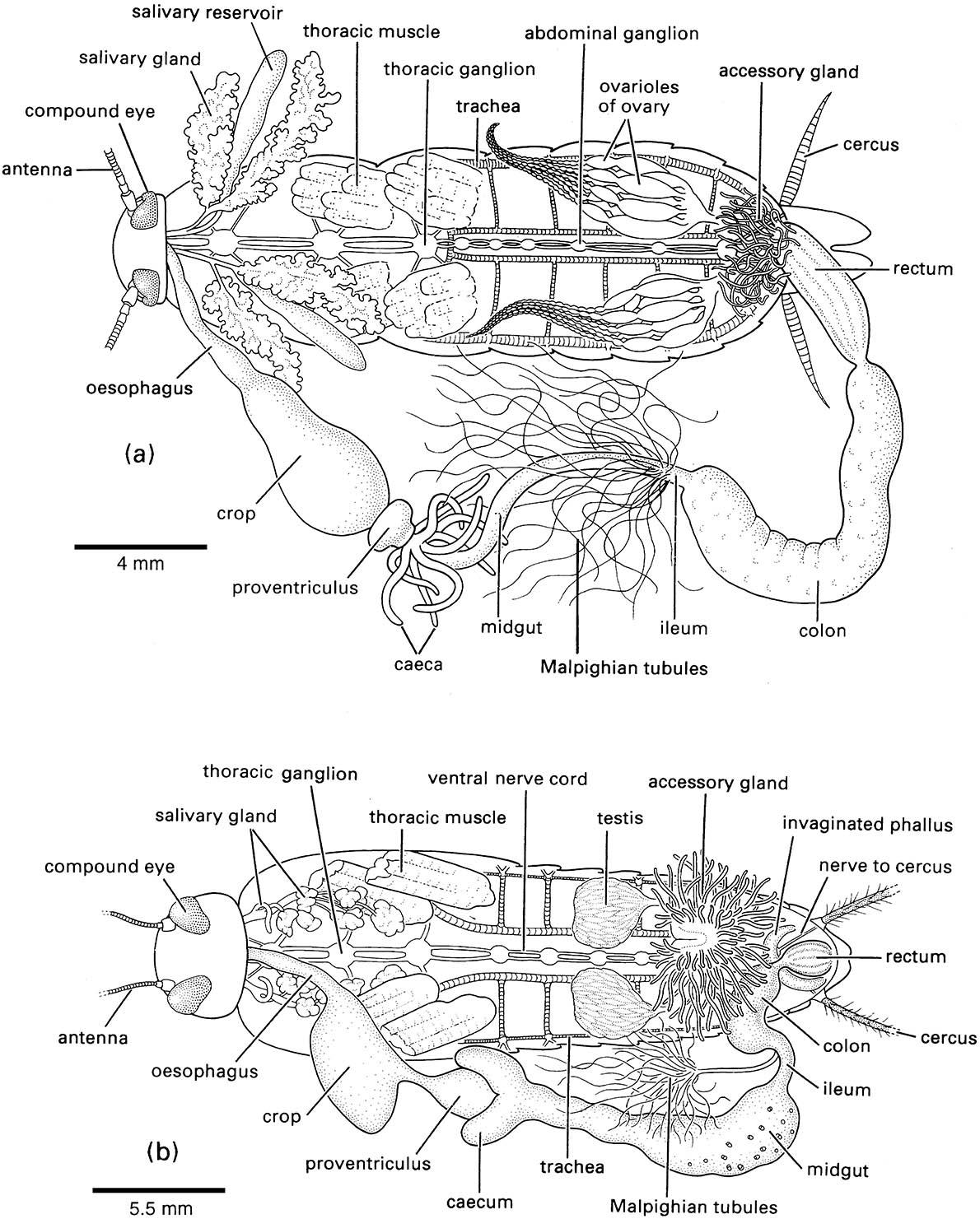
The fat body and most of the tracheae have been removed; most details of the nervous system are not shown.

The cuticular lining of the foregut and hindgut are indicated by thicker black lines. (After Dow 1986)
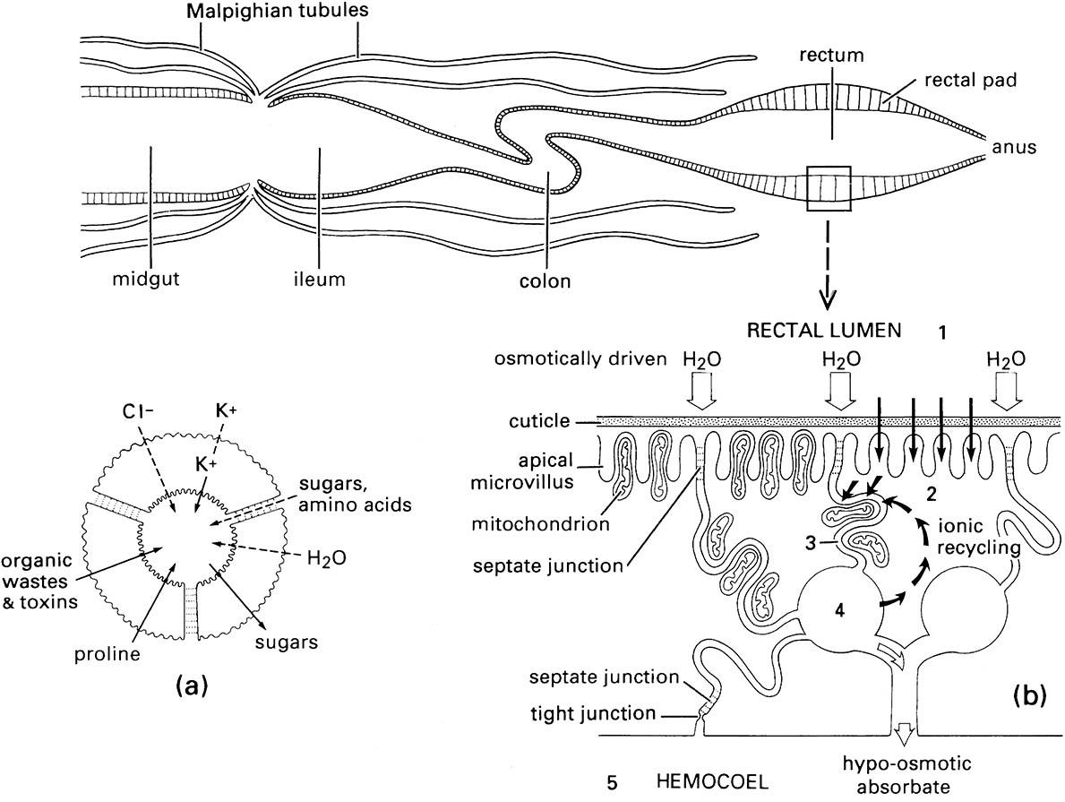
Only a few of the > 100 Malpighian tubules are drawn. (a) Transverse section of one Malpighian tubule showing probable transport of ions, water, and other substances between the surrounding hemolymph and the tubule lumen; active processes are indicated by solid arrows and passive processes by dashed arrows. (b) Diagram illustrating the movements of solutes and water in the rectal pad cells during fluid resorption from the rectal lumen. Pathways of water movement are represented by open arrows and solute movements by black arrows. Ions are actively transported from the rectal lumen (compartment 1) to the adjacent cell cytoplasm (compartment 2) and then to the intercellular spaces (compartment 3). Mitochondria are positioned to provide the energy for this active ion transport. Fluid in the spaces is hyperosmotic ( higher ion concentration) to the rectal lumen and draws water by osmosis from the lumen via the septate junctions between the cells. Water thus moves from compartment 1 to 3 to 4 and finally to 5, the hemolymph in the hemocoel. (After Bradley 1985)

