3.5. The tracheal system and gas exchange
In common with all aerobic animals, insects must obtain oxygen from their environment and eliminate carbon dioxide respired by their cells. This is gas exchange, distinguished from respiration, which strictly refers to oxygen-consuming, cellular metabolic processes. In almost all insects, gas exchange occurs by means of internal air-filled tracheae. These tubes branch and ramify through the body (Fig. 3.10). The finest branches contact all internal organs and tissues, and are especially numerous in tissues with high oxygen requirements. Air usually enters the tracheae via spiracular openings that are positioned laterally on the body, primitively with one pair per post-cephalic segment. No extant insect has more than 10 pairs (two thoracic and eight abdominal) (Fig. 3.11a), most have eight or nine, and some have one (Fig. 3.11c), two, or none (Fig. 3.11d-f ). Typically, spiracles (Fig. 3.10a) have a chamber, or atrium, with an opening-and-closing mechanism, or valve, either projecting externally or at the inner end of the atrium. In the latter type, a filter apparatus sometimes protects the outer opening. Each spiracle may be set in a sclerotized cuticular plate called a peritreme.
The tracheae are invaginations of the epidermis and thus their lining is continuous with the body cuticle. The characteristic ringed appearance of the tracheae seen in tissue sections (as in Fig. 3.7) is due to the spiral ridges or thickenings of the cuticular lining, the taenidia, which allow the tracheae to be flexible but resist compression (analogous to the function of the ringed hose of a vacuum cleaner). The cuticular linings of the tracheae are shed with the rest of the exoskeleton when the insect molts. Usually even the linings of the finest branches of the tracheal system are shed at ecdysis but linings of the fluid-filled blind endings, the tracheoles, may or may not be shed. Tracheoles are less than 1 µm in diameter and closely contact the respiring tissues (Fig. 3.10b), sometimes indenting into the cells that they supply. However, the tracheae that supply oxygen to the ovaries of many insects have very few tracheoles, the taenidia are weak or absent, and the tracheal surface is evaginated as tubular spirals projecting into the hemolymph. These aptly named aeriferous tracheae have a highly permeable surface that allows direct aeration of the surrounding hemolymph from tracheae that may exceed 50 µm in diameter.
In terrestrial and many aquatic insects the tracheae open to the exterior via the spiracles (an open tracheal system) (Fig. 3.11a-c). In contrast, in some aquatic and many endoparasitic larvae spiracles are absent (a closed tracheal system) and the tracheae divide peripherally to form a network. This covers the general body surface (allowing cutaneous gas exchange) (Fig. 3.11d) or lies within specialized filaments or lamellae (tracheal gills) (Fig. 3.11e,f ). Some aquatic insects with an open tracheal system carry gas gills with them (e.g. bubbles of air); these may be temporary or permanent (section 10.3.4).
The volume of the tracheal system ranges between 5% and 50% of the body volume depending on species and stage of development. The more active the insect, the more extensive is the tracheal system. In many insects, parts of tracheae are dilated or enlarged to increase the reservoir of air, and in some species the dilations form air sacs (Fig. 3.11b), which collapse readily because the taenidia of the cuticular lining are reduced or absent. Sometimes the tracheal volume may decrease within a developmental stage as air sacs are occluded by growing tissues. Air sacs reach their greatest development in very active flying insects, such as bees and cyclorrhaphous Diptera. They may assist flight by increasing buoyancy, but their main function is in ventilation of the tracheal system.
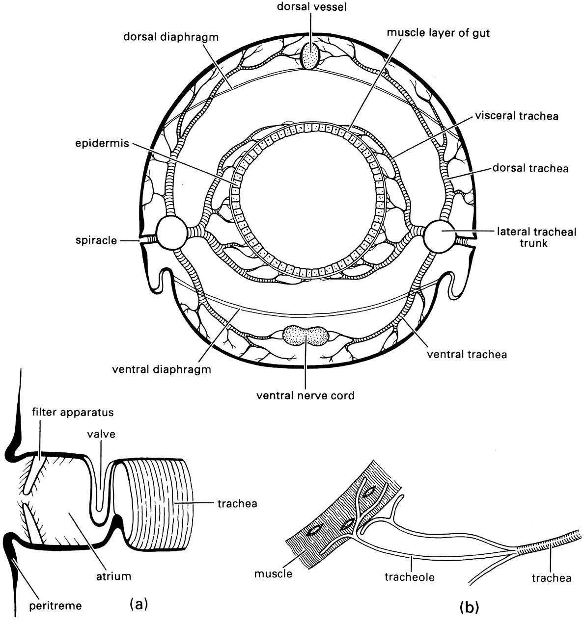
Enlargements show: (a) an atriate spiracle with closing valve at inner end of atrium; (b) tracheoles running to a muscle fiber. (After Snodgrass 1935)
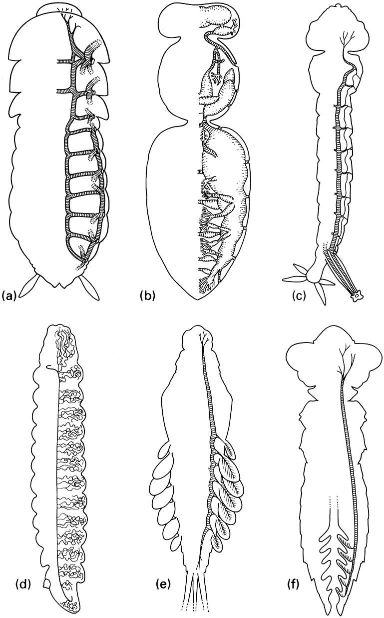
(a) Simple tracheae with valved spiracles, as in cockroaches. (b) Tracheae with mechanically ventilated air sacs, as in honey bees. (c) Metapneustic system with only terminal spiracles functional, as in mosquito larvae. (d) Entirely closed tracheal system with cutaneous gas exchange, as in most endoparasitic larvae. (e) Closed tracheal system with abdominal tracheal gills, as in mayfly nymphs. (f) Closed tracheal system with rectal tracheal gills, as in dragonfly nymphs. (After Wigglesworth 1972; details in (a) after Richards & Davies 1977, ( b) after Snodgrass 1956, (c) after Snodgrass 1935, (d) after Wigglesworth 1972)