3. Internal anatomy and physiology
What you see if you dissect open the body of an insect is a complex and compact masterpiece of functional design. Figure 3.1 shows the “insides” of two omnivorous insects, a cockroach and a cricket, which have relatively unspecialized digestive and reproductive systems. The digestive system, which includes salivary glands as well as an elongate gut, consists of three main sections. These function in storage, biochemical breakdown, absorption, and excretion. Each gut section has more than one physiological role and this may be reflected in local structural modifications, such as thickening of the gut wall or diverticula (extensions) from the main lumen. The reproductive systems depicted in Fig. 3.1 exemplify the female and male organs of many insects. These may be dominated in males by very visible accessory glands, especially as the testes of many adult insects are degenerate or absent. This is because the spermatozoa are produced in the pupal or penultimate stage and stored. In gravid female insects, the body cavity may be filled with eggs at various stages of development, thereby obscuring other internal organs. Likewise, the internal structures (except the gut) of a well-fed, late-stage caterpillar may be hidden within the mass of fat body tissue.
The insect body cavity, called the hemocoel (haemocoel) and filled with fluid hemolymph (haemolymph), is lined with endoderm and ectoderm. It is not a true coelom, which is defined as a mesoderm-lined cavity. Hemolymph (so-called because it combines many roles of vertebrate blood (hem/haem) and lymph) bathes all internal organs, delivers nutrients, removes metabolites, and performs immune functions. Unlike vertebrate blood, hemolymph rarely has respiratory pigments and therefore has little or no role in gaseous exchange. In insects this function is performed by the tracheal system, a ramification of air-filled tubes (tracheae), which sends fine branches throughout the body. Gas entry to and exit from tracheae is controlled by sphincter-like structures called spiracles that open through the body wall. Non-gaseous wastes are filtered from the hemolymph by filamentous Malpighian tubules (named after their discoverer), which have free ends distributed through the hemocoel. Their contents are emptied into the gut from which, after further modification, wastes are eliminated eventually via the anus.
All motor, sensory, and physiological processes in insects are controlled by the nervous system in con- junction with hormones (chemical messengers). The brain and ventral nerve cord are readily visible in dissected insects, but most endocrine centers, neurosecretion sites, numerous nerve fibers, muscles, and other tissues cannot be seen by the unaided eye.
This chapter describes insect internal structures and their functions. Topics covered are the muscles and locomotion (walking, swimming, and flight), the nervous system and co-ordination, endocrine centers and hormones, the hemolymph and its circulation, the tracheal system and gas exchange, the gut and digestion, the fat body, nutrition and microorganisms, the excretory system and waste disposal, and lastly the reproductive organs and gametogenesis. A full account of insect physiology cannot be provided in one chapter, and we direct readers to Chapman (1998) for a comprehensive treatment, and to relevant chapters in the Encyclopedia of Insects (Resh & Cardé 2003).
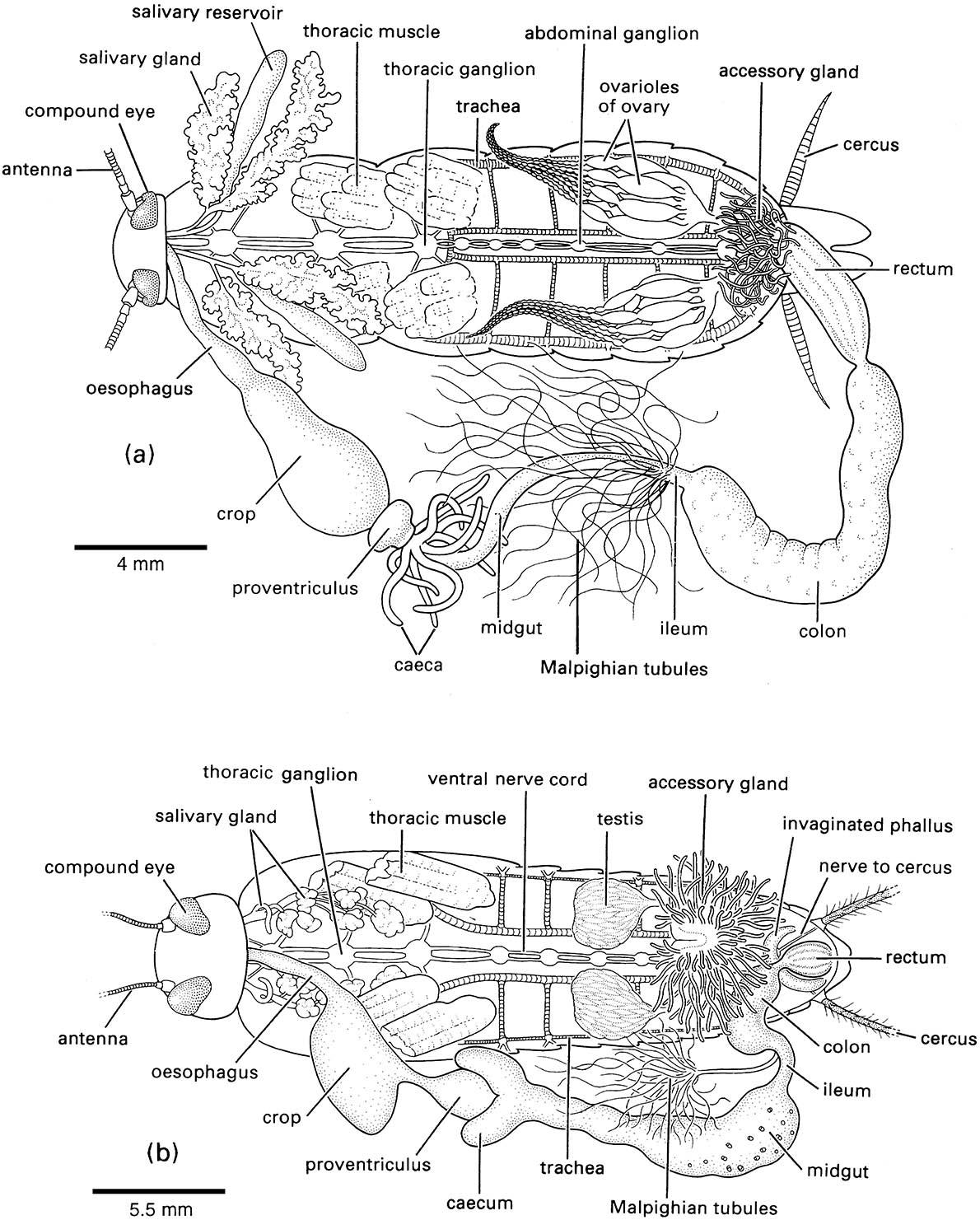
The fat body and most of the tracheae have been removed; most details of the nervous system are not shown.

