3.1.1. Muscles
Vertebrates and many non-insect invertebrates have striated and smooth muscles, but insects have only striated muscles, so-called because of overlapping thicker myosin and thinner actin filaments giving a microscopic appearance of cross-banding. Each striated muscle fiber comprises many cells, with a common plasma membrane and sarcolemma, or outer sheath. The sarcolemma is invaginated, but not broken, where an oxygen-supplying tracheole (section 3.5, Fig. 3.10b) contacts the muscle fiber. Contractile myofibrils run the length of the fiber, arranged in sheets or cylinders. When viewed under high magnification, a myofibril comprises a thin actin filament sandwiched between a pair of thicker myosin filaments. Muscle contraction involves the sliding of filaments past each other, stimulated by nerve impulses. Innervation comes from one to three motor axons per bundle of fibers, each separately tracheated and referred to as one muscle unit, with several units grouped in a functional muscle.
There are several different muscle types. The most important division is between those that respond synchronously, with a contraction cycle once per impulse, and fibrillar muscles that contract asynchronously, with multiple contractions per impulse. Examples of the latter include some flight muscles (see below) and the tymbal muscle of cicadas (section 4.1.4).
There is no inherent difference in action between muscles of insects and vertebrates, although insects can produce prodigious muscular feats, such as the leap of a flea or the repetitive stridulation of the cicada tympanum. Reduced body size benefits insects because of the relationship between (i) power, which is proportional to muscle cross-section and decreases with reduction in size by the square root, and (ii) the body mass, which decreases with reduction in size by the cube root. Thus the power : mass ratio increases as body size decreases.
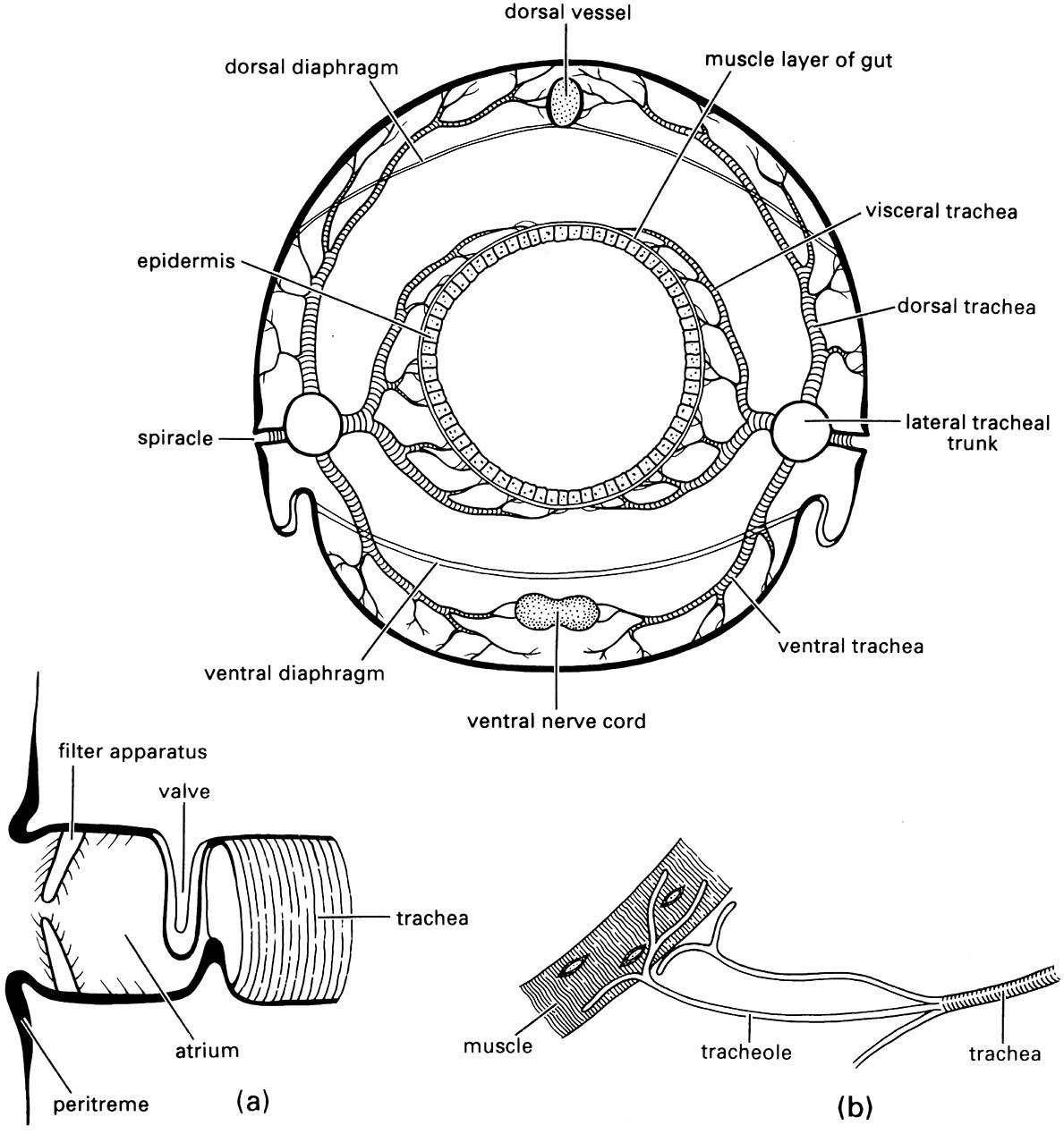
Enlargements show: (a) an atriate spiracle with closing valve at inner end of atrium; (b) tracheoles running to a muscle fiber. (After Snodgrass 1935)

