3.2. The nervous system and co-ordination
The complex nervous system of insects integrates a diverse array of external sensory and internal physiological information and generates some of the behaviors discussed in Chapter 4. In common with other animals, the basic component is the nerve cell, or neuron (neurone), composed of a cell body with two projections (fibers) — the dendrite, which receives stimuli; and the axon, which transmits information, either to another neuron or to an effector organ such as a muscle. Insect neurons release a variety of chemicals at synapses to either stimulate or inhibit effector neurons or muscles. In common with vertebrates, particularly important neurotransmitters include acetylcholine and catecholamines such as dopamine. Neurons (Fig. 3.5) are of at least four types:
- sensory neurons receive stimuli from the insect’s environment and transmit them to the central nervous system (see below);
- interneurons (or association neurons) receive information from and transmit it to other neurons;
- motor neurons receive information from inter- neurons and transmit it to muscles;
- neuroendocrine cells (section 3.3.1).
The cell bodies of interneurons and motor neurons are aggregated with the fibers interconnecting all types of nerve cells to form nerve centers called ganglia. Simple reflex behavior has been well studied in insects (described further in section 4.5), but insect behavior can be complex, involving integration of neural information within the ganglia.
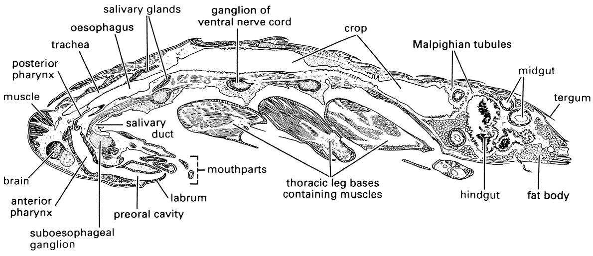
The central nervous system (CNS) (Fig. 3.6) is the principal division of the nervous system and consists of series of ganglia joined by paired longitudinal nerve cords called connectives. Primitively there are a pair of ganglia per body segment but usually the two ganglia of each thoracic and abdominal segment are fused into a single structure and the ganglia of all head segments are coalesced to form two ganglionic centers — the brain and the suboesophageal (subesophageal) ganglion (seen in Fig. 3.7). The chain of thoracic and abdominal ganglia found on the floor of the body cavity is called the ventral nerve cord. The brain, or the dorsal ganglionic center of the head, is composed of three pairs of fused ganglia (from the first three head segments):
- protocerebrum, associated with the eyes and thus bearing the optic lobes;
- deutocerebrum, innervating the antennae;
- tritocerebrum, concerned with handling the signals that arrive from the body.
Coalesced ganglia of the three mouthpart-bearing segments form the suboesophageal ganglion, with nerves emerging that innervate the mouthparts.
The visceral (or sympathetic) nervous system consists of three subsystems — the stomodeal (or stomatogastric) (which includes the frontal ganglion); the ventral visceral; and the caudal visceral. Together the nerves and ganglia of these subsystems innervate the anterior and posterior gut, several endocrine organs (corpora cardiaca and corpora allata), the reproductive organs, and the tracheal system including the spiracles.
The peripheral nervous system consists of all of the motor neuron axons that radiate to the muscles from the ganglia of the CNS and stomodeal nervous system plus the sensory neurons of the cuticular sensory structures (the sense organs) that receive mechanical, chemical, thermal, or visual stimuli from an insect’s environment. Insect sensory systems are discussed in detail in Chapter 4.
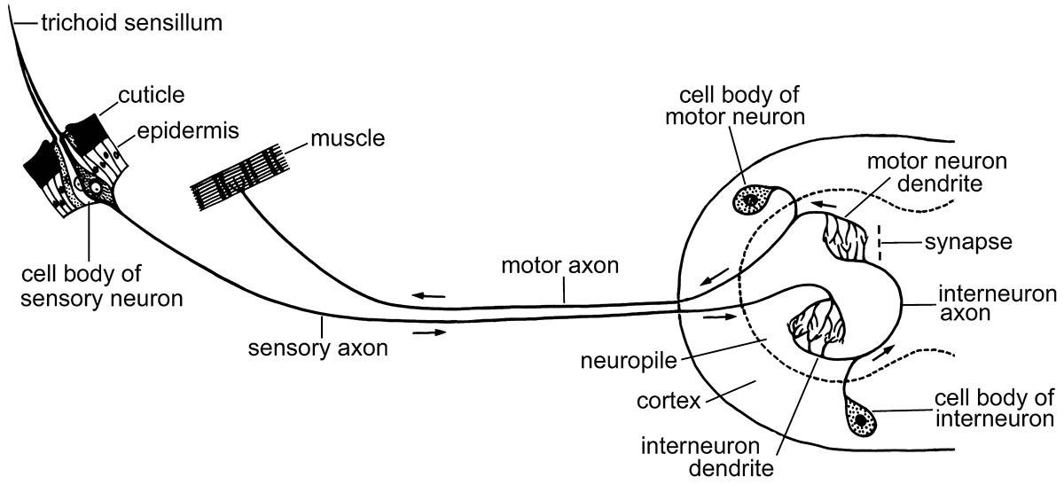
The arrows show the paths of nerve impulses along nerve fibers (axons and dendrites). The ganglion, with its outer cortex and inner neuropile, is shown on the right. (After various sources)

Varying degrees of fusion of ganglia occur from the least to the most specialized: (a) three separate thoracic and eight abdominal ganglia, as in Dictyopterus (Coleoptera: Lycidae) and Pulex (Siphonaptera: Pulicidae); (b) three thoracic and six abdominal, as in Blatta (Blattodea: Blattidae) and Chironomus (Diptera: Chironomidae); (c) two thoracic and considerable abdominal fusion of ganglia, as in Crabro and Eucera (Hymenoptera: Crabronidae and Anthophoridae); (d) highly fused with one thoracic and no abdominal ganglia, as in Musca , Calliphora, and Lucilia ( Diptera: Muscidae and Calliphoridae); (e) extreme fusion with no separate suboesophageal ganglion, as in Hydrometra (Hemiptera: Hydrometridae) and Rhizotrogus (Scarabaeidae). (After Horridge 1965)