2.4. The thorax
The thorax is composed of three segments: the first or prothorax, the second or mesothorax, and the third or metathorax. Primitively, and in apterygotes (bristletails and silverfish) and immature insects, these segments are similar in size and structural complexity. In most winged insects the mesothorax and metathorax are enlarged relative to the prothorax and form a pterothorax, bearing the wings and associated musculature. Wings occur only on the second and third segments in extant insects although some fossils have prothoracic winglets (Fig. 8.2) and homeotic mutants may develop prothoracic wings or wing buds. Almost all nymphal and adult insects have three pairs of thoracic legs — one pair per segment. Typically the legs are used for walking, although various other functions and associated modifications occur (section 2.4.1). Openings (spiracles) of the gas-exchange, or tracheal, system (section 3.5) are present laterally on the second and third thoracic segments at most with one pair per segment. However, a secondary condition in some insects is for the mesothoracic spiracles to open on the prothorax.
The tergal plates of the thorax are simple structures in apterygotes and in many immature insects, but are variously modified in winged adults. Thoracic terga are called nota (singular: notum), to distinguish them from the abdominal terga. The pronotum of the prothorax may be simple in structure and small in comparison with the other nota, but in beetles, mantids, many bugs, and some Orthoptera the pronotum is expanded and in cockroaches it forms a shield that covers part of the head and mesothorax. The pterothoracic nota each have two main divisions — the anterior wing-bearing alinotum and the posterior phragma-bearing postnotum (Fig. 2.18). Phragmata (singular: phragma) are plate-like apodemes that extend inwards below the antecostal sutures, marking the primary intersegmental folds between segments; phragmata provide attachment for the longitudinal flight muscles (Fig. 2.7d). Each alinotum (sometimes confusingly referred to as a “notum”) may be traversed by sutures that mark the position of internal strengthening ridges and commonly divide the plate into three areas — the anterior prescutum, the scutum, and the smaller posterior scutellum.
The lateral pleural sclerites are believed to be derived from the subcoxal segment of the ancestral insect leg (Fig. 8.4a). These sclerites may be separate, as in silverfish, or fused into an almost continuous sclerotic area, as in most winged insects. In the pterothorax, the pleuron is divided into two main areas — the anterior episternum and the posterior epimeron — by an internal pleural ridge, which is visible externally as the pleural suture (Fig. 2.18); the ridge runs from the pleural coxal process (which articulates with the coxa) to the pleural wing process (which articulates with the wing), providing reinforcement for these articulation points. The epipleurites are small sclerites beneath the wing and consist of the basalaria anterior to the pleural wing process and the posterior subalaria, but often reduced to just one basalare and one subalare, which are attachment points for some direct flight muscles. The trochantin is the small sclerite anterior to the coxa.
The degree of ventral sclerotization on the thorax varies greatly in different insects. Sternal plates, if present, are typically two per segment: the eusternum and the following intersegmental sclerite or intersternite (Fig. 2.7c), commonly called the spinasternum (Fig. 2.18) because it usually has an internal apodeme called the spina (except for the metasternum which never has a spinasternum). The eusterna of the prothorax and mesothorax may fuse with the spinasterna of their segment. Each eusternum may be simple or divided into separate sclerites — typically the presternum, basisternum, and sternellum. The eusternum may be fused laterally with one of the pleural sclerites and is then called the laterosternite. Fusion of the sternal and pleural plates may form precoxal and postcoxal bridges (Fig. 2.18).
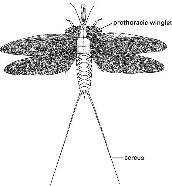
(After Kukalová 1970)
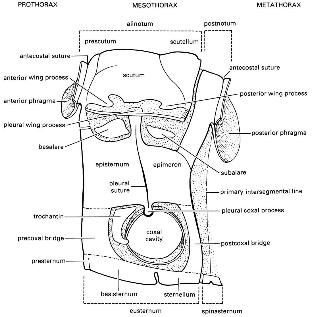
(After Snodgrass 1935)
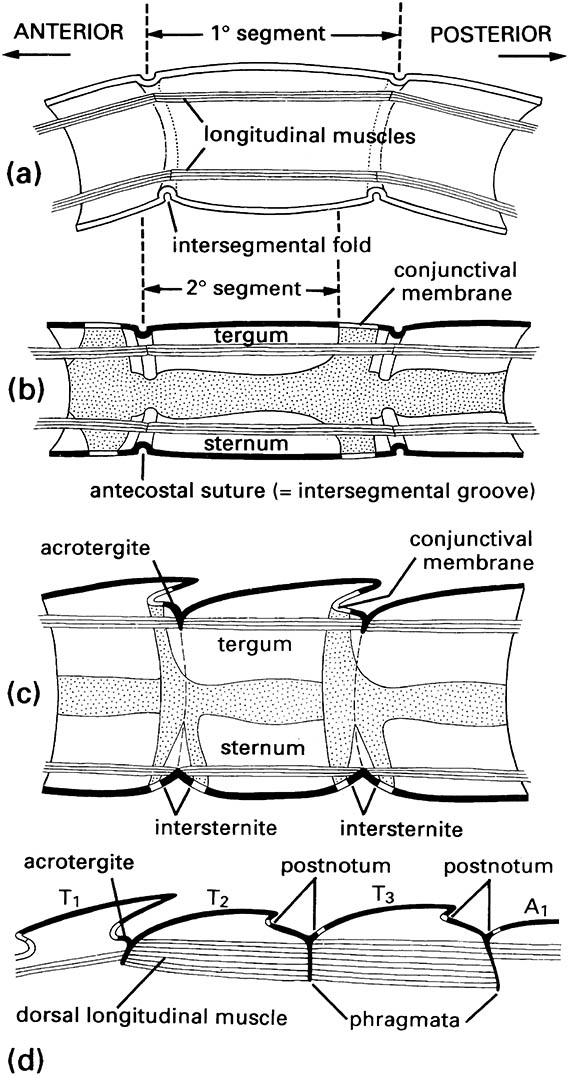
(a) Primary segmentation, as seen in soft-bodied larvae of some insects. (b) Simple secondary segmentation. (c) More derived secondary segmentation. (d) Longitudinal section of dorsum of the thorax of winged insects, in which the acrotergites of the second and third segments have enlarged to become the postnota. (After Snodgrass 1935)
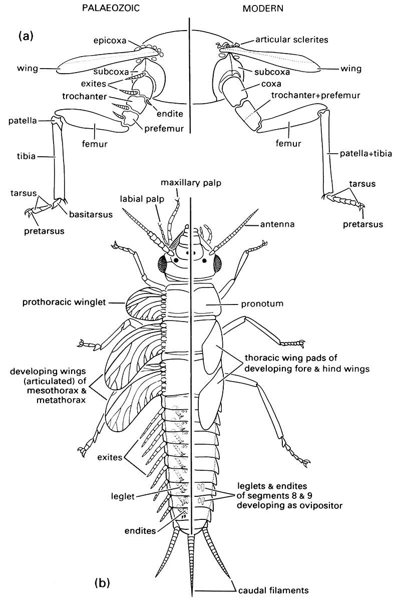
(a) thoracic segment of adult showing generalized condition of appendages; (b) dorsal view of nymphal morphology. (Modified from Kukalová-Peck 1991; to incorporate ideas of J.W.H. Trueman (unpublished))

