2.2. Segmentation and tagmosis
Metameric segmentation, so distinctive in annelids, is visible only in some unsclerotized larvae (Fig. 2.7a). The segmentation seen in the sclerotized adult or nymphal insect is not directly homologous with that of larval insects, as sclerotization extends beyond each primary segment (Fig. 2.7b,c). Each apparent segment represents an area of sclerotization that commences in front of the fold that demarcates the primary segment and extends almost to the rear of that segment, leaving an unsclerotized area of the primary segment, the conjunctival or intersegmental membrane. This secondary segmentation means that the muscles, which are always inserted on the folds, are attached to solid rather than to soft cuticle. The apparent segments of adult insects, such as on the abdomen, are secondary in origin, but we refer to them simply as segments throughout this text.
In adult and nymphal insects, and hexapods in general, one of the most striking external features is the amalgamation of segments into functional units. This process of tagmosis has given rise to the familiar tagmata (regions) of head, thorax, and abdomen. In this process the 20 original segments have been divided into an embryologically detectable six-segmented head, three-segmented thorax, and 11-segmented abdomen (plus primitively the telson), although varying degrees of fusion mean that the full complement is never visible.
Before discussing the external morphology in more detail, some indication of orientation is required. The bilaterally symmetrical body may be described according to three axes:
- longitudinal, or anterior to posterior, also termed cephalic (head) to caudal (tail);
- dorsoventral, or dorsal (upper) to ventral (lower);
- transverse, or lateral (outer) through the longitudinal axis to the opposite lateral (Fig. 2.8).
For appendages, such as legs or wings, proximal or basal refers to near the body, whereas distal or apical means distant from the body. In addition, structures are mesal, or medial, if they are nearer to the midline (median line), or lateral if closer to the body margin, relative to other structures.
Four principal regions of the body surface can be recognized: the dorsum or upper surface; the venter or lower surface; and the two lateral pleura (singular: pleuron), separating the dorsum from the venter and bearing limb bases, if these are present. Sclerotization that takes place in defined areas gives rise to plates called sclerites. The major segmental sclerites are the tergum (the dorsal plate; plural: terga), the sternum (the ventral plate; plural: sterna), and the pleuron (the side plate). If a sclerite is a subdivision of the tergum, sternum, or pleuron, the diminutive terms tergite, sternite, and pleurite may be applied.
The abdominal pleura are often at least partly membranous, but on the thorax they are sclerotized and usually linked to the tergum and sternum of each segment. This fusion forms a box, which contains the leg muscle insertions and, in winged insects, the flight muscles. With the exception of some larvae, the head sclerites are fused into a rigid capsule. In larvae (but not nymphs) the thorax and abdomen may remain membranous and tagmosis may be less apparent (such as in most wasp larvae and fly maggots) and the terga, sterna, and pleura are rarely distinct.
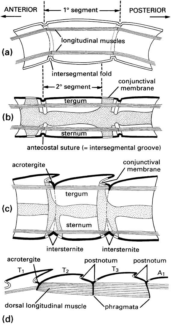
(a) Primary segmentation, as seen in soft-bodied larvae of some insects. (b) Simple secondary segmentation. (c) More derived secondary segmentation. (d) Longitudinal section of dorsum of the thorax of winged insects, in which the acrotergites of the second and third segments have enlarged to become the postnota. (After Snodgrass 1935)
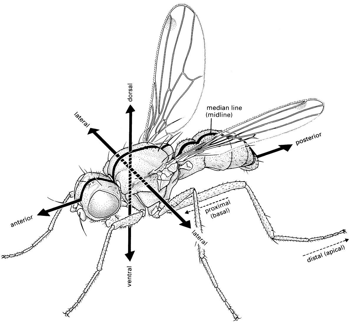
(After McAlpine 1987)

