2.3. The head
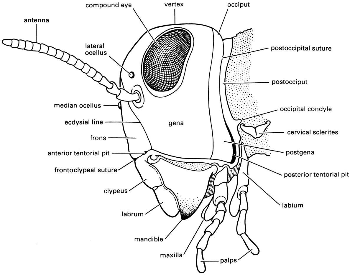
The rigid cranial capsule has two openings, one posteriorly through the occipital foramen to the prothorax, the other to the mouthparts. Typically the mouthparts are directed ventrally (hypognathous), although sometimes anteriorly (prognathous) as in many beetles, or posteriorly (opisthognathous) as in, for example, aphids, cicadas, and leafhoppers. Several regions can be recognized on the head (Fig. 2.9): the posterior horse-shoe-shaped posterior cranium (dorsally the occiput) contacts the vertex dorsally and the genae (singular: gena) laterally; the vertex abuts the frons anteriorly and more anteriorly lies the clypeus, both of which may be fused into a frontoclypeus. In adult and nymphal insects, paired compound eyes lie more or less dorsolaterally between the vertex and genae, with a pair of sensory antennae placed more medially. In many insects, three light-sensitive “simple” eyes, or ocelli, are situated on the anterior vertex, typically arranged in a triangle, and many larvae have stemmatal eyes.
The head regions are often somewhat weakly delimited, with some indications of their extent coming from sutures (external grooves or lines on the head). Three sorts may be recognized:
- remnants of original segmentation, generally restricted to the postoccipital suture;
- ecdysial lines of weakness where the head capsule of the immature insect splits at molting (section 6.3), including an often prominent inverted “Y”, or epicranial suture, on the vertex (Fig. 2.10); the frons is delimited by the arms (also called frontal sutures) of this “Y”;
- grooves that reflect the underlying internal skeletal ridges, such as the frontoclypeal or epistomal suture, which often delimits the fronts from the more anterior clypeus.
The head endoskeleton consists of several invaginated ridges and arms (apophyses, or elongate apodemes), the most important of which are the two pairs of tentorial arms, one pair being posterior, the other anterior, sometimes with an additional dorsal component. Some of these arms may be absent or, in pterygotes, fused to form the tentorium, an endoskeletal strut. Pits are discernible on the surface of the cranium at the points where the tentorial arms invaginate. These pits and the sutures may provide prominent landmarks on the head but usually they bear little or no association with the segments.
The segmental origin of the head is most clearly demonstrated by the mouthparts (section 2.3.1). From anterior to posterior, there are six fused head segments:
- labral;
- antennal, with each antenna equivalent to an entire leg;
- postantennal, fused with the antennal segment;
- mandibular;
- maxillary;
- labial.
The neck is mainly derived from the first part of the thorax and is not a segment.
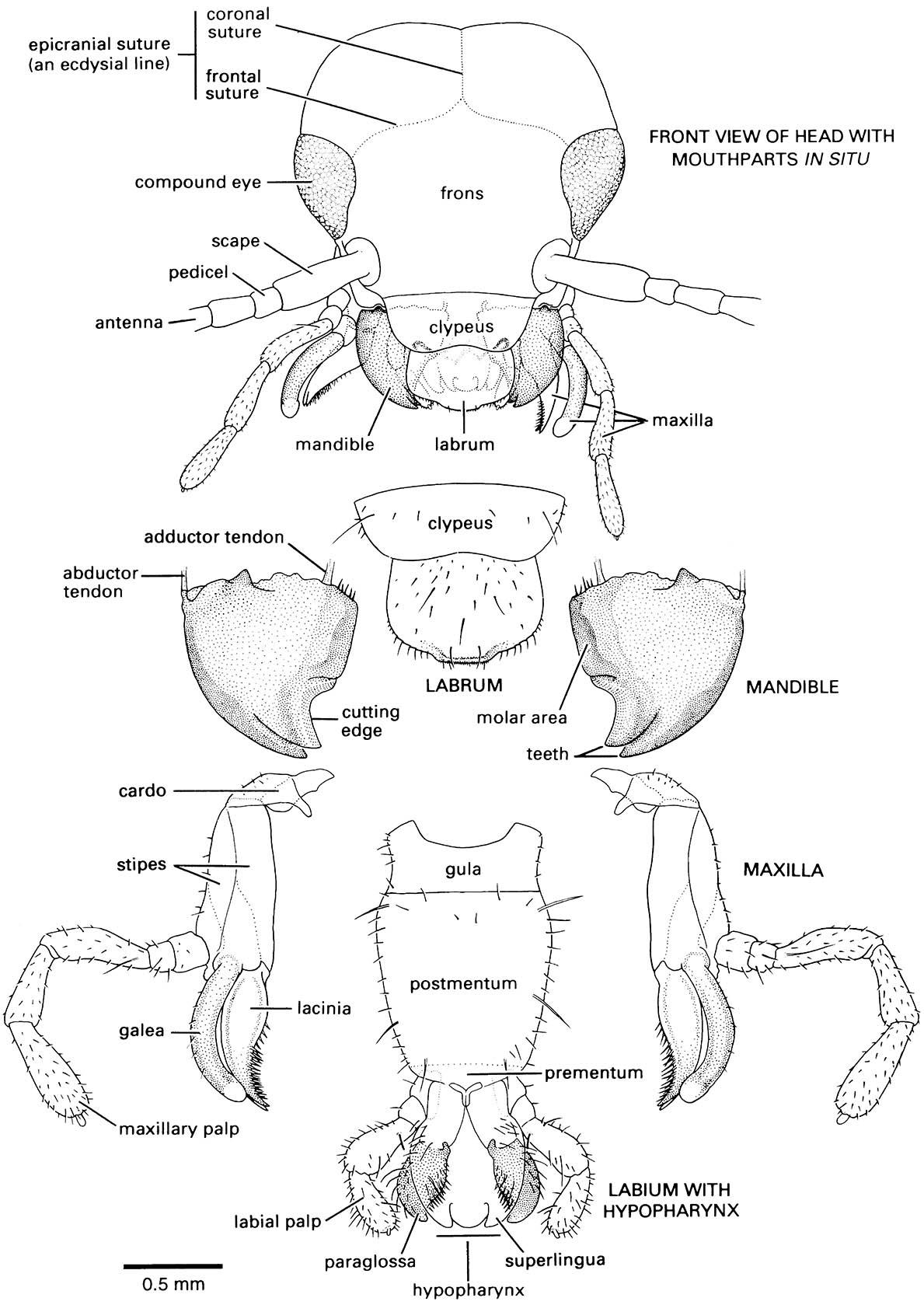
Note that the head is prognathous and thus a gular plate, or gula, occurs in the ventral neck region.

