4.4.2. Stemmata
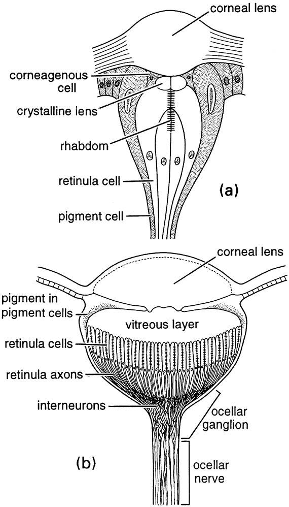
The only visual organs of larval holometabolous insects are stemmata, sometimes called larval ocelli (Fig. 4.9a). These organs are located on the head, and vary from a single pigmented spot on each side to six or seven larger stemmata, each with numerous photoreceptors and associated nerve cells. In the simplest stemma, a cuticular lens overlies a crystalline body secreted by several cells. Light is focused by the lens onto a single rhabdom. Each stemma points in a different direction so that the insect sees only a few points in space according to the number of stemmata.
Some caterpillars increase the field of view and fill in the gaps between the direction of view of adjacent stemmata by scanning movements of the head. Other larvae, such as those of sawflies and tiger beetles, possess more sophisticated stemmata. They consist of a two-layered lens that forms an image on an extended retina composed of many rhabdoms, each receiving light from a different part of the image. In general, stemmata seem designed for high light sensitivity, with resolving power relatively low.

(a) a simple stemma of a lepidopteran larva; (b) a light-adapted median ocellus of a locust. ((a) After Snodgrass 1935; (b) after Wilson 1978)

