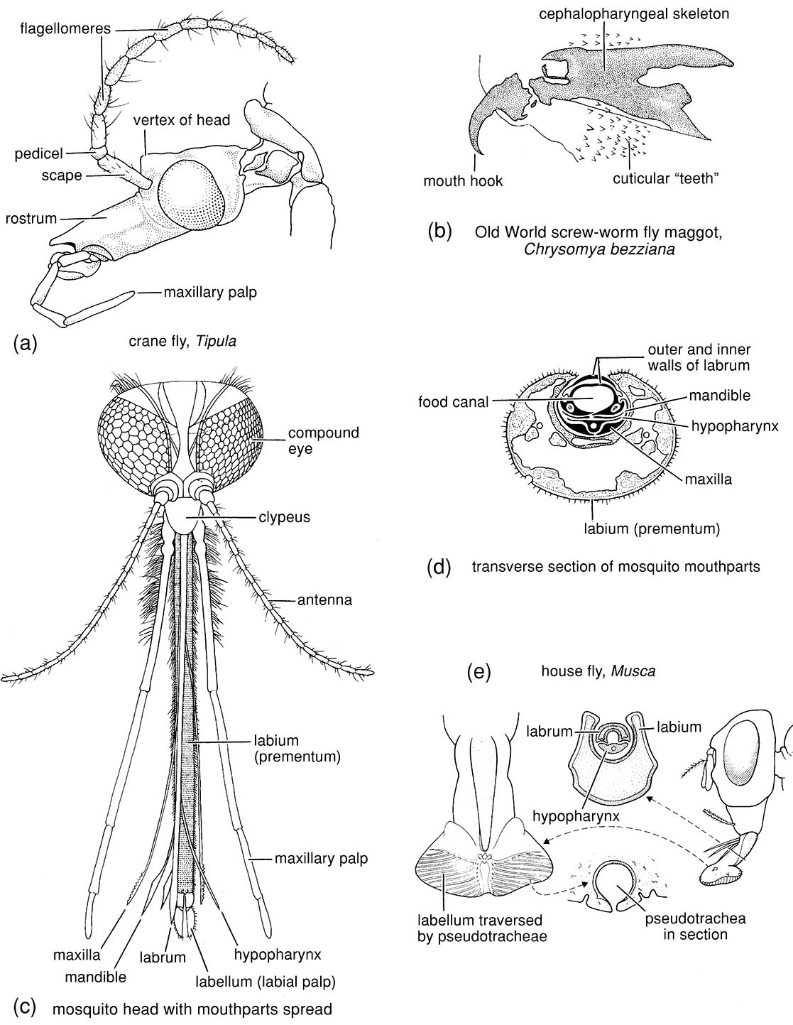
Box 15.5. Diptera (flies)
The Diptera is an order containing perhaps some 125,000 described species, in roughly 130 families, with several thousands of species of medical or veterinary importance. Development is holometabolous, and adults have variously modified mouthparts, mesothoracic wings, and metathoracic halteres (balancers) (Fig. 2.22f ). The larvae lack true legs (Fig. 6.6h), and their head structure ranges from a complete sclerotized capsule to acephaly with no external capsule and only an internal skeleton. The pupae are adecticous and obtect, or exarate in a puparium (Fig. 6.7e,f).
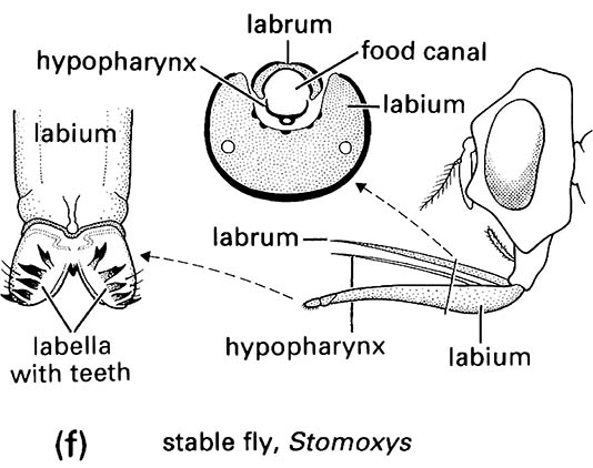
The paraphyletic Nematocera comprises crane flies, mosquitoes, midges, gnats, and relatives; these have slender antennae with upwards of six flagellomeres and a three- to five-segmented maxillary palp (illustrated for a crane fly (Tipulidae: Tipula) in (a), after McAlpine 1981). Brachycera contains heavier-built flies including hover flies, bee flies, dung flies, and blow flies. They have more solid, often shorter antennae with fewer (less than seven) flagellomeres, often with terminal arista (Fig. 2.17i); the maxillary palps have only one or two segments (illustrated for Muscidae in (e) and (f )). Within the Brachycera, schizophoran Cyclorrhapha use a ptilinum to aid emergence from the puparium.
Fly larvae have a wide variety of habits. Many nematoceran larvae are aquatic (Box 10.5), and brachyceran larvae show a phylogenetic radiation into drier and more specialized larval habits, including phytophagy, predation and parasitization of other arthropods, and myiasis-induction in vertebrates (section 15.3). Myiasis- inducing maggots have a much reduced head but with sclerotized mouthparts known as mouth hooks (illustrated for a third-instar larva of the Old World screw-worm fly, Chrysomya bezziana (Calliphoridae) in (b), after Ferrar 1987), which scrape the living flesh of the host.
Adult dipteran mouthparts are illustrated in frontal view (c) (after Freeman & Bracegirdle 1971) and trans- verse section (d) (after Jobling 1976) for a female mosquito. All dipterans typically have a tubular sucking organ, the proboscis, comprising elongate mouthparts (usually including the labrum). A biting-and-sucking type of proboscis appears to be a primitive dipteran feature. Although biting functions have been lost and regained with modifications more than once, blood-feeding is frequent, and leads to the importance of the Diptera as vectors of disease. The blood-feeding female nematocerans — Culicidae (mosquitoes); Ceratopogonidae (biting midges); Psychodidae: Phlebotominae (sand flies); and Simuliidae (black flies) — have generally similar mouthparts, but differ in proboscis length, allowing penetration of the host to different depths. Mosquitoes can probe deep in search of capillaries, but other blood-feeding nematocerans operate more superficially where a pool of blood is induced in the wound. The labium ends in two sensory labella (singular: labellum), forming a protective sheath for the functional mouthparts. Enclosed are serrate-edged, cutting mandibles and maxillary lacinia, the curled labrum—epipharynx, and the hypopharynx, all of which are often termed stylets. When feeding, the labrum, mandibles, and laciniae act as a single unit driven through the skin of the host. The flexible labium remains bowed outside the wound. Saliva, which may contain anticoagulant, is injected through a salivary duct that runs the length of the sharply pointed and often toothed hypopharynx. Blood is transported up a food canal formed from the curled labrum sealed by either the paired mandibles or the hypopharynx. Capillary blood can flow unaided, but blood must be sucked or pumped from a pool with pumping action from two muscular pumps: the cibarial located at the base of the food canal, and the pharyngeal in the pharynx between the cibarium and midgut.
Many mouthparts are lost in the “higher” flies, and the remaining mouthparts are modified for lapping food using pseudotracheae of the labella as “sponges” (illustrated for a house fly (Muscidae: Musca) in (e), after Wigglesworth 1964). With neither mandibles nor maxillary lacinia to make a wound, blood-feeding cyclorrhaphans often use modified labella, in which the inner surfaces are adorned with sharp teeth (illustrated for a stable fly (Muscidae: Stomoxys) in (f ), after Wigglesworth 1964). Through muscular contraction and relaxation, the labellar lobes dilate and contract repeatedly, creating an often painful rasping of the labellar teeth to give a pool of blood. The hypopharynx applies saliva which is dissipated via the labellar pseudotracheae. Uptake of blood is via capillary action through “food furrows” lying dorsal to the pseudotracheae, with the aid of three pumps operating synchronously to produce continuous suction from labella to pharynx. A prelabral pump produces the contractions in the labella, with a more proximal labral pump linked via a feeding tube to the cibarial pump.
The mouthparts and their use in feeding have implications for disease transmission. Shallow-feeding species such as black flies are more involved in trans- mission of microfilariae, such as those of Onchocerca, which aggregate just beneath the skin, whilst deeper feeders such as mosquitoes transmit pathogens that circulate in the blood. The transmission from fly to host is aided by the introduction of saliva into the wound, and many parasites aggregate in the salivary glands or ducts. Filariae, in contrast, are too large to enter the wound through this route, and leave the insect host by rupturing the labium or labella during feeding.
Phylogenetic relationships are considered in section 7.4.2 and depicted in Fig. 7.6.
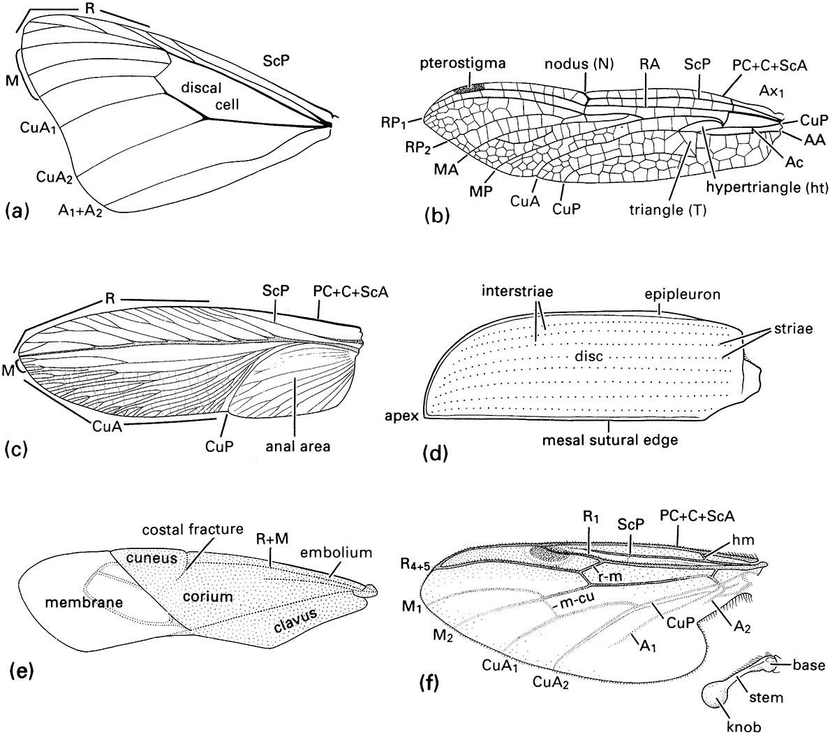
(a) fore wing of a butterfly of Danaus (Lepidoptera: Nymphalidae); (b) fore wing of a dragonfly of Urothemis (Odonata: Anisoptera: Libellulidae); (c) fore wing or tegmen of a cockroach of Periplaneta (Blattodea: Blattidae); (d) fore wing or elytron of a beetle of Anomala (Coleoptera: Scarabaeidae); (e) fore wing or hemelytron of a mirid bug (Hemiptera: Heteroptera: Miridae) showing three wing areas — the membrane, corium, and clavus; (f ) fore wing and haltere of a fly of Bibio (Diptera: Bibionidae) (after J.W.H. Trueman, unpublished. ((a-d) After Youdeowei 1977; (f) after McAlpine 1981)
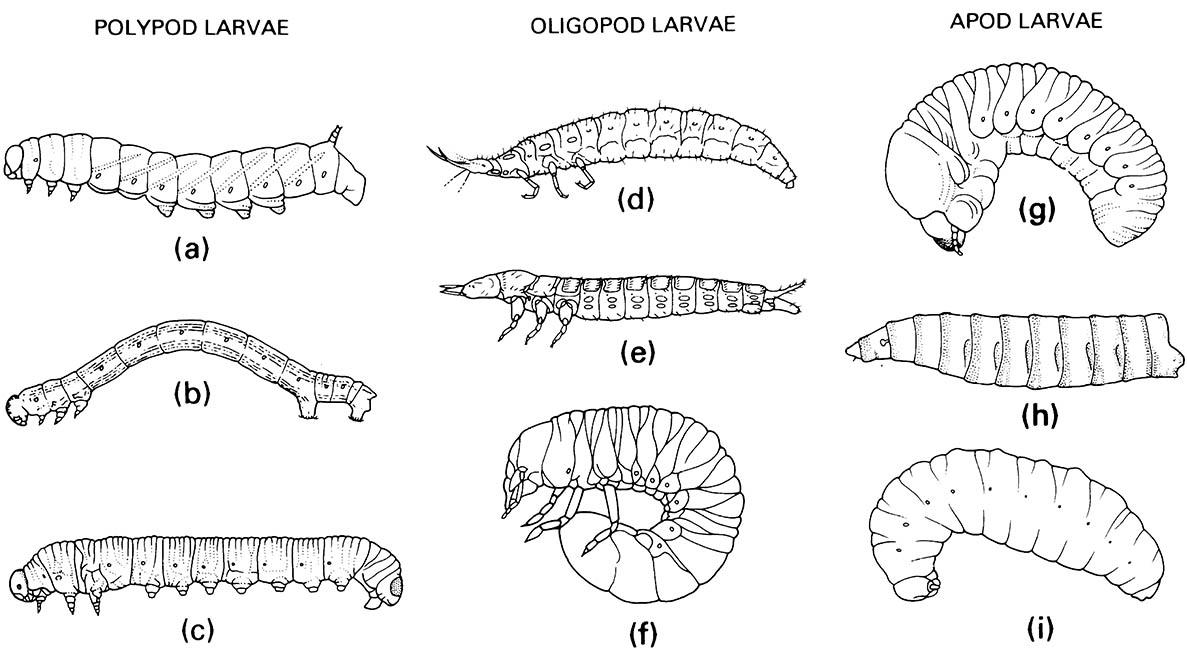
(a) Lepidoptera: Sphingidae; (b) Lepidoptera: Geometridae; (c) Hymenoptera: Diprionidae. Oligopod larvae: (d) Neuroptera: Osmylidae; (e) Coleoptera: Carabidae; (f ) Coleoptera: Scarabaeidae. Apod larvae: (g) Coleoptera: Scolytidae; (h) Diptera: Calliphoridae; (i) Hymenoptera: Vespidae. ((a, e-g) After Chu 1949; (b, c) after Borror et al. 1989; (h) after Ferrar 1987; (i) after CSIRO 1970)
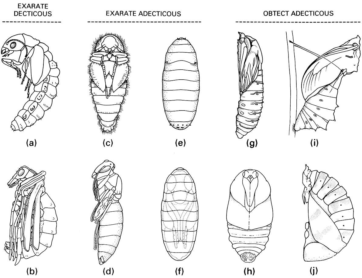
Exarate decticous pupae: (a) Megaloptera: Sialidae; (b) Mecoptera: Bittacidae. Exarate adecticous pupae: (c) Coleoptera: Dermestidae; (d) Hymenoptera: Vespidae; (e,f ) Diptera: Calliphoridae, puparium and pupa within. Obtect adecticous pupae: (g) Lepidoptera: Cossidae; (h) Lepidoptera: Saturniidae; (i) Lepidoptera: Papilionidae, chrysa lis; (j) Coleoptera: Coccinellidae. ((a) After Evans 1978; (b, c, e, g) after CSIRO 1970; (d) after Chu 1949; (h) after Common 1990; (i) after Common & Waterhouse 1972; (j) after Palmer 1914)
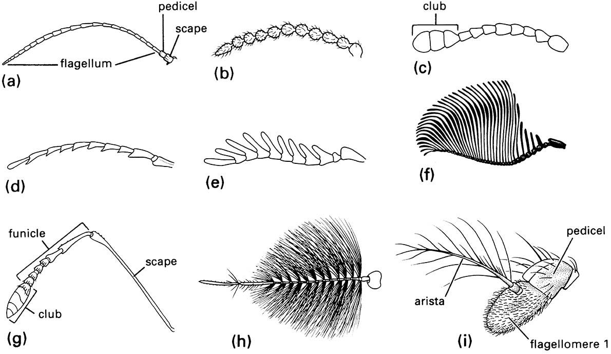
(a) filiform — linear and slender; (b) moniliform — like a string of beads; (c) clavate or capitate — distinctly clubbed; (d) serrate — saw-like; (e) pectinate — comb-like; (f ) flabellate — fan-shaped; (g) geniculate — elbowed; (h) plumose — bearing whorls of setae; and (i) aristate — with enlarged third segment bearing a bristle.
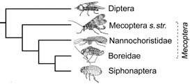
The broken lines indicate a paraphyletic taxon, with its name italicized; s. str. refers to the restricted sense. (After Whiting 2002)


