Figures 2.3
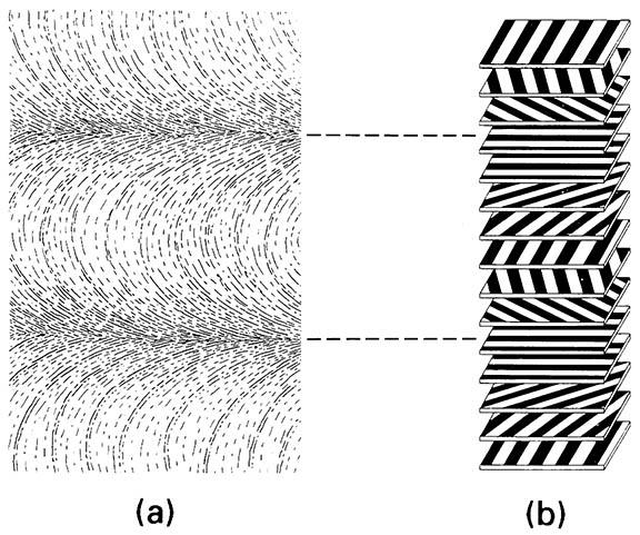
Figures 2.3. The ultrastructure of cuticle (from a transmission electron micrograph).
The arrangement of chitin microfibrils in a helicoidal array produces characteristic (though artifactual) parabolic patterns. (b) Diagram of how the rotation of microfibrils produces a lamellar effect owing to microfibrils being either aligned or non-aligned to the plane of sectioning. (After Filshie 1982)

