Figures 3.18
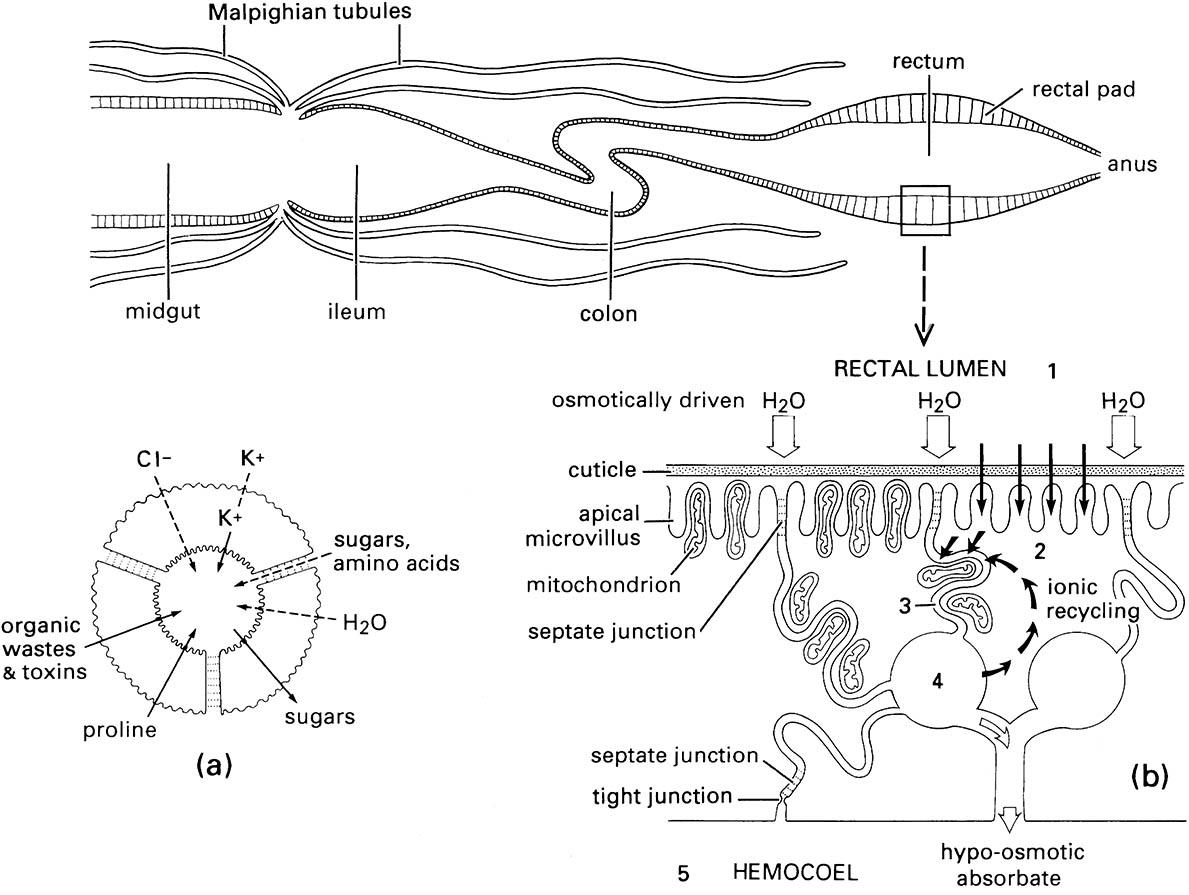
Only a few of the > 100 Malpighian tubules are drawn. (a) Transverse section of one Malpighian tubule showing probable transport of ions, water, and other substances between the surrounding hemolymph and the tubule lumen; active processes are indicated by solid arrows and passive processes by dashed arrows. (b) Diagram illustrating the movements of solutes and water in the rectal pad cells during fluid resorption from the rectal lumen. Pathways of water movement are represented by open arrows and solute movements by black arrows. Ions are actively transported from the rectal lumen (compartment 1) to the adjacent cell cytoplasm (compartment 2) and then to the intercellular spaces (compartment 3). Mitochondria are positioned to provide the energy for this active ion transport. Fluid in the spaces is hyperosmotic ( higher ion concentration) to the rectal lumen and draws water by osmosis from the lumen via the septate junctions between the cells. Water thus moves from compartment 1 to 3 to 4 and finally to 5, the hemolymph in the hemocoel. (After Bradley 1985)

