Amoebae
The two best-studied insect amoebae are Malpighamoeba mellificae and Malamoeba locustae, which are associated with the honeybee, Apis mellifera, and the Melanoplus grasshoppers, respectively. The cysts of the honeybee amoeba are ingested and excyst, releasing slender primary trophozoites that penetrate and multiply in the midgut epithelium. Secondary trophozoites emerge from these cells and migrate to the lumen of the Malpighian tubules. These trophozoites, having pseudopodia, feed in the lumen and cause a flattening of the epithelial layer and a distension of the tubules. The brush border in contact with the amoeba swells in size and loses the associated secretory transport vesicles. Infected tubules contain a mix of secondary trophozoites, precysts, and cysts. The primary damage to the host bee is the malfunction of the Malpighian tubules. Both numbers of amoeba and the presence of other disease agents determine the severity of the amoebiasis in the bee. In general, this disease either induces stress or under appropriate conditions in the springtime can be debilitative, resulting in hive dwindling.
Malamoeba locustae, also known as Malamoeba locusta, has been detected in a wide range of grasshopper species and in a single Thysanuran species. Its life cycle is very similar to that observed with M. mellificae. The host grasshoppers ingest the resistant uninucleate cysts, and excysted primary trophozoites invade the midgut and caecal tissues. Within these tissues the trophozoites grow and divide, and within about 10 days release progeny secondary trophozoites into the lumen. These cells migrate to the lumen of the Malpighian tubules and undergo additional cell divisions (Fig. 45).
The vegetative development of this amoeba damages the serosal membrane of the tubules, inhibiting their response to insect diuretic hormone. The infected tubules become packed with trophozoites and cysts. At high levels, M. locustae may inhibit the excretory function of the tubules and cause the grasshoppers to become lethargic prior to death. The distended, amoebainfected tubules may rupture, releasing both trophozoites and cysts into the hemocoel. These amoebas are quickly recognized as non-self and are encapsulated by circulating phagocytic hemocytes. This disease, although a problem in laboratory cultured grasshoppers, is rarely detected in natural populations.
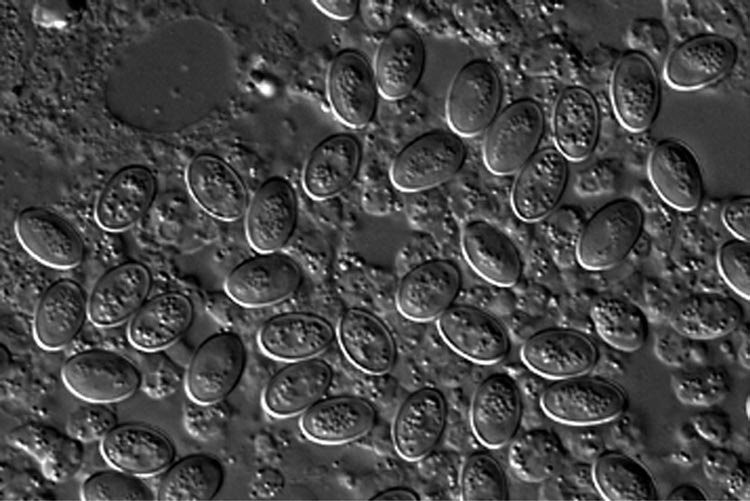
Figure 45 Light micrograph of the cysts of Malamoeba locusta released from infected Malpighian tubules.

American Tropical Silkworm Moths


